



Abstract
Background
Ovarian hormones may contribute to the vulnerability to depression as well as to the response to antidepressants (ADs). Previously we reported that acute systemic treatment with estradiol or progesterone blocked the ability of the selective serotonin reuptake inhibitor (SSRI), fluvoxamine, to inhibit serotonin transporter (SERT) function in ovariectomized (OVX) rats. In this study, behavioral consequences as well as receptor mechanisms underlying these hormonal effects were investigated.
Methods
Using the forced swimming test (FST), the acute effect of estradiol and/or progesterone on fluvoxamine’s AD-like effects was investigated. Using in vivo chronoamperometry, the effect of local application of estradiol or progesterone into the CA3 region of the hippocampus of OVX rats on 5-HT clearance as well as on the ability of fluvoxamine to slow 5-HT clearance was investigated.
Results
The decreased immobility and increased swimming caused by fluvoxamine in the FST was blocked in rats treated with estradiol and/or progesterone. Local application of estradiol, but not progesterone, slowed 5-HT clearance and both hormones blocked the ability of fluvoxamine to slow 5-HT clearance. Use of hormone receptor agonists, antagonists and hormone-BSA complexes revealed that the effects of estradiol are mediated by activation of membrane as well as nuclear estrogen receptors (ER). The AD-like effect of estradiol involved ERβ and GPR30 whereas its blockade of fluvoxamine’s effects was ERα-mediated. The effects of progesterone occurred solely by activation of intracellular progesterone receptors.
Conclusion
Targeting of ERβ or GPR30 might reveal a strategy to permit beneficial effects of estrogen without its deleterious effect on SSRI-efficacy.
Keywords: estradiol, progesterone, FST, chronoamperometry, serotonin transporter, SSRI
INTRODUCTION
Depression is more common in females than males (1) and in women it may be more common at times of falling estradiol levels (2). Women tend to report more depressive and dysphoric symptoms, even if not full-blown major depressive disorder (MDD), during times of large hormonal changes, including menopause, the premenstrual period and postpartum which suggest that hormonal changes are playing a role in these symptoms (3, 4). Nevertheless, the effectiveness on depressed mood of hormone replacement therapy during the postmenopausal period is somewhat controversial and both positive (5) and negative (6) results have been reported.
In ovariectomized rats, we have shown that acute systemic administration of either estradiol benzoate (EB) and/or progesterone (P) blocked the ability of SSRIs, fluvoxamine and citalopram, to inhibit the function of what is widely considered their initial cellular target in brain – the serotonin transporter (SERT) (7). In addition, when given systemically, EB, but not P, blocked the function of the SERT (8).
The behavioral consequences of these hormonal effects are not known. The most widely-used behavioral test in rats that is predictive of antidepressant efficacy in patients is the forced swimming test (FST) (9, 10). AD-like effects of estradiol and progesterone have been shown in the FST (11-13). However, few studies have examined if ovarian steroids alter the effects of ADs in these tests. In the first part of this study, the effects of acute ovarian hormones on the ability of fluvoxamine to decrease immobility in the FST were examined.
A major advance in understanding estrogen’s actions was the discovery and cloning in 1996 of a second estrogen receptor (ER), ERβ (14), that is genetically distinct from the ERα subtype first cloned ten years earlier (15). Recently, a novel membrane-associated ER, GPR30, was identified (16, 17). GPR30 is a seven transmembrane-spanning G-protein coupled receptor that promotes rapid estrogen signaling in a variety of cell types (18). GPR30 shows specific high affinity binding to estradiol and related estrogens (19). A second part of this study examined the mechanisms underlying the effects of ovarian hormones on SERT function and on the ability of fluvoxamine to block SERT function, using in vivo chronoamperometry after local application of hormones, hormone receptor-subtype specific agonists and antagonists. The use of local application provides more specificity for mechanistic studies as both hormone and drug effects are primarily confined to the area into which they are administered. All in vivo chronoamperometry experiments were carried out in the CA3 region of the hippocampus as under our experimental conditions, the active clearance of 5-HT in this area is mediated primarily by the SERT (20). Further, there is evidence for the involvement of the hippocampus in depression and the effects of antidepressants (21). Finally, both intracellular and membrane ERs in the hippocampus can mediate hippocampal effects of estrogen (22).
METHODS AND MATERIALS
Ovariectomized (OVX) rats (Sprague-Dawley; 250-350g, Harlan, Houston, TX) were housed on a 12:12h light/dark cycle with lights on at 0700. All animal procedures were in accordance with the National Institutes of Health Guide for the Care and Use of Laboratory Animals and were approved by the local Institutional Animal Care and Use Committee. OVX rats were used 2-3 weeks after surgery.
Forced swimming test (FST)
The FST was carried out as described (23, 24) except the pretest and test sessions were separated by about 75 hours rather than the usual 24 hours. Rats were placed individually in Pyrex cylinders (21 × 46 cm) that were filled with water to a 30-cm depth. The rats were removed 15 min later, dried and placed into their home cage. Either 24 or 75 hours after their first exposure, the animals were again placed in the swim apparatus for 5 minutes and behaviors monitored from above by video camera for subsequent analysis. Fluvoxamine (Sigma-Aldrich, St Louis, MO) or saline was administered subcutaneously three times at 1, 5 and 23.5 hours prior to the test session. The behavioral rater was blind with respect to the experimental conditions being scored. A time sampling technique was employed whereby the predominant behavior in each 5-s period of the 300-s test was recorded. Climbing behavior consists of upward directed movements of the forepaws along the side of the swim chamber. Swimming behavior is defined as movement (usually horizontal) throughout the swim chamber, which also includes crossing into another quadrant. Immobility is assigned when no additional activity is observed other than that required to keep the rat’s head above the water.
The schedule for hormones and fluvoxamine administration is shown schematically in Figure 1.
Figure 1.
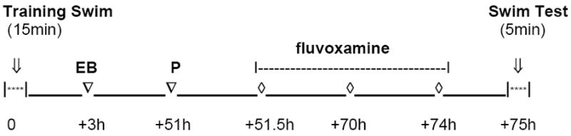
Timeline for administration of hormones (EB and/or P) and/or fluvoxamine treatment in the FST.
Hormone treatments
OVX rats were treated subcutaneously with either EB (25μg, in 100μl peanut oil, 74-75h prior to the experiment; Sigma-Aldrich, St Louis, MO), P (0.5mg, in 100μl peanut oil, 24h prior to the experiment; Sigma-Aldrich, St Louis, MO), or the combination of EB/P, or with vehicle (peanut oil), 24 or 74-75h prior to the experiment.
These doses of hormones produce serum concentrations similar to those found in proestrus (8). For each hormone treatment, 12 rats were used, six of which received fluvoxamine and the other six received saline injections.
In vivo chronoamperometry
This was carried out essentially as described previously (10, 25-27).
Animal preparation
OVX rats were anesthetized with chloralose (70mg/Kg)/urethane (700mg/kg) administered intraperitoneally (ip), after tracheal intubation and placed into a stereotaxic apparatus (David Kopf instruments). The body temperature of the rat was maintained between 37-38°C using a water-circulated heating pad. Rectal temperature was continuously monitored with a probe attached to a YSI temperature meter. The scalp was incised and reflected and a hole drilled in the skull at the desired coordinates. A small burr hole was drilled over the posterior cortex for placement of Ag/AgCl reference electrode.
Electrode preparation
Carbon fiber electrodes (30μm tip diameter, 95-175μm in length) were coated with Nafion to improve the selectivity of the electrode, then tested for sensitivity to 5-hydroxyindole-3-acetic acid (5-HIAA, 250μM, Sigma-Aldrich, St Louis, MO) and calibrated in vitro with 5-HT. We only used electrodes displaying a selectivity ratio for 5-HT over 5-HIAA > than 1000:1 and a linear response (r2 >0.977) to 5-HT (0.5-3.0μM).
Micropipette preparation
The carbon fiber electrode was positioned adjacent a multibarrel micropipette (4 or 7 barrels) using a micromanipulator and the tip separation, determined using a dissecting microscope, was between 250-350 μm. The electrode and multibarrel micropipette were then attached using sticky wax. Micropipette barrels were filled with 5-HT (200 μM), fluvoxamine (400μM) [fluvoxamine is always used at 4x the amount of 5-HT applied], and the other drugs to be tested. All drugs were prepared in 0.1M phosphate buffered saline (PBS) and supplemented with 100 μM ascorbic acid. The pH of all solutions was 7.4. All drugs were delivered by pressure ejection in a volume of 20-100nl, using a PLI-100 reproducible pico-injector. The volume of the fluid was determined using a dissection microscope (Nikon SMZ-1) fitted with a reticule eye piece.
Electrochemical recordings
The electrode-pipette assembly was lowered into the CA3 region of hippocampus (stereotaxic coordinates (mm) anterior-posterior (AP), -4.10 from bregma; mediolateral (ML), +3.30 from midline; dorsal-ventral (DV), -3.60 from dura (28). Chronoamperometric recordings were started 20-30min after the lowering of the assembly to allow the baseline electrochemical signal to stabilize. High-speed chronoamperometric recordings were made using the Fast-12 and Fast-16 systems (Quanteon LLC, Nicholasville, KY). Oxidation potentials of 100msec pulses of +0.55 V versus Ag/AgCl were delivered one per second; the electrode was held at the resting potential of 0.0 V between measurements. Oxidation and reduction currents were digitally integrated during the last 80msec of each 100msec voltage pulse. Analyzed in this study is the clearance time parameter T80, the time it takes for the peak signal amplitude to be reduced by 80%.
Hormones, hormone receptor antagonists and receptor subtype-selective agonists tested were, 17β-estradiol (E2), progesterone (P), RU486 (Sigma-Aldrich, St Louis, MO), ICI 182,780, propyl-pyrazoltriol (PPT) and tetrahydrochrysene (THC), diarylpropionitrile (DPN), G1 (Tocris, Ellisville, MO), and Progesterone 3-CMO:BSA (P-BSA; Steraloids, Inc.Newport, RI); they were dissolved in PBS or PBS containing ethanol (less than 0.0001%).
The doses of hormones were selected based on our previous study (8); the lowest doses that induced changes in SERT function were selected. The doses of ER agonists were based on the affinity of the agonist compared to that of E2 for ER subtypes (29, 30) and on pilot studies. 5-12 rats were used for each tested drug in each of the two experimental designs 1) time course of the effects of drugs on 5-HT clearance; 2) time course of the effects of drugs on fluvoxamine’s blockade of 5-HT clearance.
Statistical analysis
Data were analyzed (SigmaStats, Systat Software Inc, San Jose, CA) by either one way or two way ANOVA followed by Newman-Keuls post hoc tests for multiple comparisons. For non-parametric analyses, Kruskal-Wallis one way analysis of variance on ranks followed by Dunns analysis for multiple comparisons was used. Only when there was a significant main effect and/or interaction effect in the ANOVA’s were post-hoc analyses carried out. Significance was determined at p < 0.05.
RESULTS
Effect of estradiol and/or progesterone on the antidepressant-like effect of fluvoxamine
EB inhibited the effect of fluvoxamine on 5-HT clearance when fluvoxamine was administered ~72h after EB (8). In order to test the effect of fluvoxamine in the FST at the same time after administration of EB and carry out the training session before EB treatment, the test session was carried out 75h after the pretest (Figure 1). Fluvoxamine (10 mg/kg) significantly decreased immobility and increased swimming behavior in control rats (Figure 2). However, these effects were blocked in rats treated with EB (25μg in 100μl peanut oil) and/or P (500μg in 100μl peanut oil). The group treated with EB alone showed some tendency for increased swimming behavior from that in the control rats; however, this increase did not reach significance.
Figure 2.
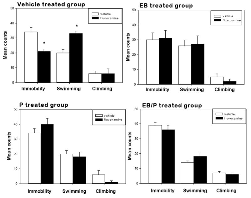
Effect of hormone treatments on fluvoxamine-induced behaviors in the FST. OVX rats were treated subcutaneously (sc) with estradiol benzoate (EB, 25μg, in 100μl peanut oil, about 75h prior to the experiment) and/or progesterone (P, 0.5mg, in 100μl peanut oil, 24h prior to the experiment) or vehicle (peanut oil, 24 or 75 h prior to the experiment). Fluvoxamine (10mg/kg) or saline were given 3 times within a 24h period, at 23.5, 5 and 1 hour before the test session. Mean counts of climbing, swimming and immobility were sampled every 5 sec during the 5 min test period. Bars and brackets represent the mean value ± SEM, n= 6/group. Two way ANOVA revealed a significant main effect for hormone treatment (F(3,51) = 4.583, p< 0.01). *p<0.02, Student-Newman-Keuls post hoc comparison of fluvoxamine’s effect with the corresponding control value for each behavior in each treatment group.
Effect of local application of estradiol on 5-HT clearance and on fluvoxamine-induced blockade of 5-HT clearance
The time course of the effect of local application of 17-β-estradiol (E2, 20pmoles) on the clearance of exogenous 5-HT (2-15pmoles) was examined from 1-120min. This time course study was carried out in order to detect membrane/intracellular receptor-mediated effects; effects occurring rapidly (1-10min) would involve membrane receptors and those occurring at later time points would involve intracellular receptors. The clearance time parameter, T80, derived from the generated 5-HT electrochemical signal, is represented in Figure 3. E2 increased T80 rapidly (within a minute) with this effect becoming significant at 10 min and persisting for up to 120min (Figure 4).
Figure 3.
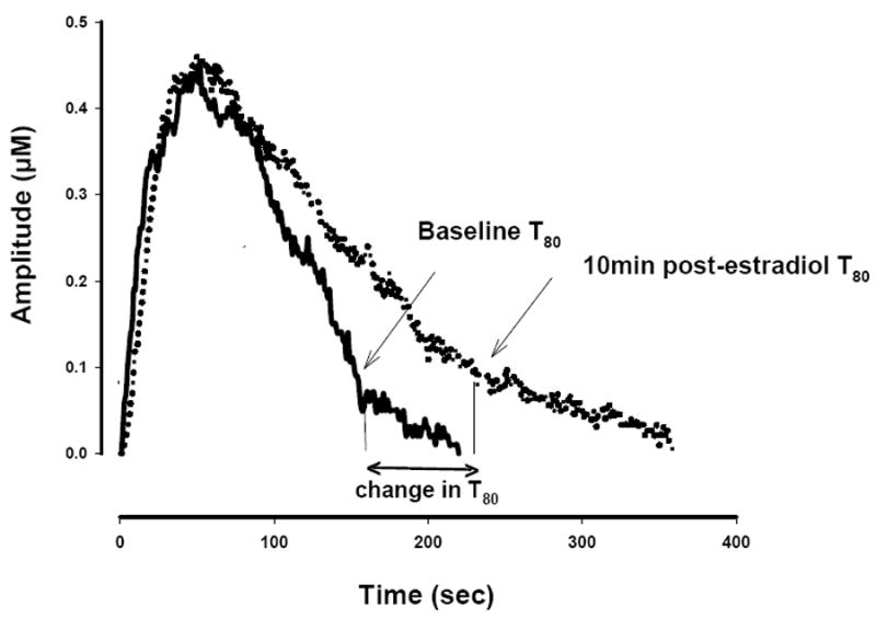
Representative 5-HT electrochemical signals illustrating the effect of locally applied 17-β estradiol into the CA3 region of the hippocampus of an OVX rat. The signal was generated by local application of 5-HT (5.2 pmoles). Estradiol 20 pmoles was pressure ejected 10min before the next application of 5-HT. Shown here is the clearance time, T80 parameter: the time it takes for the peak signal amplitude to be reduced by 80%. For clarity, only oxidation signals are shown.
Figure 4.
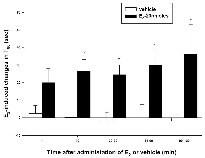
Time course of the effect of local application of E2 on the 5-HT clearance time parameter, T80. The time course was examined from 1 to 120 min after local application of E2 (20 pmoles) or PBS as the control vehicle. For these experiments, the same amount of 5-HT was pressure-ejected in a given rat at all-time points after zero. Bars and brackets represent the increase from baseline level in T80 values (in seconds) after the application of E2, ± SEM (n=4-5). Two way ANOVA revealed a significant effect of hormone (F(1,47) = 30.076, p<0.001). *p<0.05, Student-Newman-Keuls post hoc tests comparing each E2 value treatment with the corresponding control value at each time point.
Previously, we found that local application of E2 mimicked the effect of systemic administration of EB in that it blocked the inhibitory effect of fluvoxamine on 5-HT clearance. This inhibitory effect began as early as 1min after application of E2 and persisted for 60 min (8). To investigate this further, we examined the ability of an antagonist at both plasma membrane and cytosolic estrogen receptors (ERs), ICI 182,780 (31), to block effects caused by local application of E2 at 10min and 40-60min either on 5-HT clearance or on the inhibitory effect of fluvoxamine on 5-HT clearance. At 10min, ICI 182,780 (60pmoles) did not block E2’s inhibitory effect on 5-HT clearance. However, in spite of the elevated T80 value showing reduced 5-HT clearance, ICI 182,780 restored the inhibitory effect of fluvoxamine as shown by a further significant increase of the T80 value (Figure 5). At 40-60min, ICI 182,780 not only blocked the inhibition by E2 of fluvoxamine’s effect on 5-HT clearance but also the inhibitory effect of E2 alone on 5-HT clearance (Figure 5).
Figure 5.
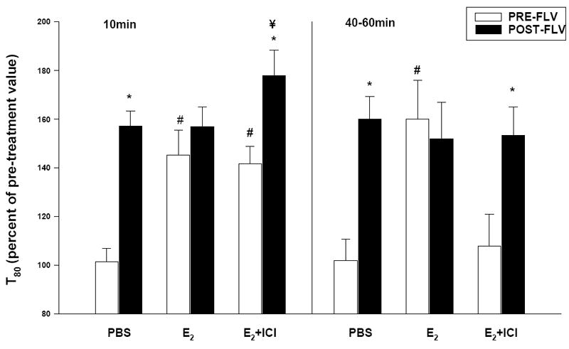
Effect of local application of E2 on 5-HT clearance and on the ability of fluvoxamine to slow clearance after ER antagonism by ICI 182,780. Pretreatment with ICI 182,780 (60 pmoles) was carried out 15min before local application of E2 (20 pmoles) and the effect on the 5-HT clearance parameter T80, as well as the effect on the ability of fluvoxamine [fluvoxamine is always used at 4x the amount of 5-HT applied] to increase T80 was measured. ICI 182,780, by itself, had no effect on basal 5-HT clearance or on the ability of fluvoxamine to slow clearance (data not shown). The clearance value prior to any PBS, fluvoxamine or hormone treatment was set at 100%. The control group was given PBS. Bar and brackets represent the T80 value as a percentage of the pre-treatment value ± SEM (n=6-10). Two way ANOVA was carried out for each time point. There was a significant main effect for fluvoxamine, both at 10min (F(1,46) = 48.336, p<0.001) and 40-60min (F(1,45) = 14.356, p<0.001) and for hormones, both E2 and ICI 182,780 + E2, at 10min (F(2,46) = 14.401, p<0.001) and 40-60min (F(2,45)=3.866, p < 0.05). A significant interaction between hormone X fluvoxamine was detected both at 10min (F(2,46) = 7.146, p <0.002) and 40-60min (F(2,45) = 5.617, p = 0.01). Student-Newman-Keuls post hoc analysis was carried out. *p<0.01, comparing the post-fluvoxamine value with the pre-fluvoxamine values in each treatment group. #p<0.001, comparing pre-fluvoxamine values in E2 or ICI 182,780 + E2 groups with the pre-fluvoxamine value in the PBS group. ¥p<0.01, comparing the fluvoxamine value in the ICI 182,780+E2 group with its effect in the other two groups.
In order to determine which ER subtypes are responsible for the effects of estradiol, the ability of specific agonists to ERα (PPT and THC, the latter also being an antagonist at ERβ; 32, 33), ERβ (DPN; 16) and to GPR30 (G1; 34) to alter SERT function or their ability to block fluvoxamine’s effects on SERT function was investigated. Similar to the effect of E2, local application of DPN (90pmoles) and G1 (0.2nmoles) increased T80 significantly whereas PPT (60pmoles) and THC (60pmoles) had no effect (Figure 6). These effects were obtained at early (1-10min) as well as at later times (40-60min). By contrast, it was PPT and THC that mimicked the ability of E2 to inhibit fluvoxamine’s effect on serotonin clearance whereas now neither DPN nor G1 had any effect. This effect was similar at early and later time points for PPT whereas THC did not block the effects of fluvoxamine at the later time (Figure 7).
Figure 6.
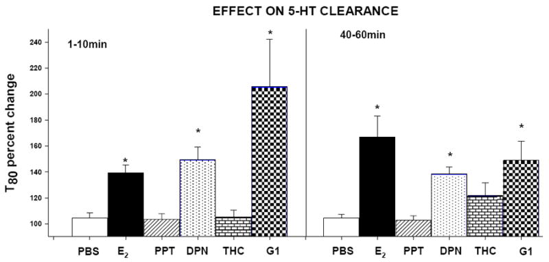
Effects of ER subtype-selective agonists on the 5-HT clearance time parameter, T80. E2 (20pmoles), PPT (ERα agonist, 60pmoles), DPN (ERβ agonist, 90pmoles), THC (agonist at ERα and also antagonist at the ERβ, 60pmoles) or G1 (GPR30 agonist, 0.2nmoles), were locally applied into the CA3 region of hippocampus and their effects were measured at an early time point (1-10min) and later time point (40-60min) post drug administration. Bar and brackets represent the T80 value as a percentage of the pre-treatment value ± SEM (n=5-12). *p<0.05, Kruskal-Wallis one way analysis of variance on ranks, followed by Dunn’s test comparing percent change in T80 values for the drugs with the percent change in T80 value in the corresponding control (PBS) group.
Figure 7.
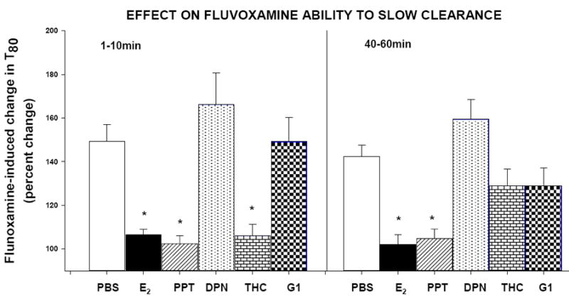
Effect of ER subtype agonists on the fluvoxamine-induced increase in the 5-HT clearance time parameter, T80. E2 (20pmoles), PPT (ERα agonist, 60pmoles), DPN (ERβ agonist, 90pmoles), THC (agonist at ERα and also antagonist at the ERβ, 60pmoles) or G1 (GPR30 agonist, 0.2nmoles), were locally applied into the CA3 region of hippocampus and their effects on the ability of fluvoxamine [used at 4x the amount of 5-HTapplied] to increase theT80 value were measured at an early time point (1-10min) and later time point (40-60min) after drug administration. Bar and brackets represent the percent change in T80 value after fluvoxamine ± SEM (n=5-12). *p<0.05, Kruskal-Wallis one way analysis of variance on ranks, followed by Dunn’s test comparing percent change in the T80 value post-fluvoxamine after E2 or the hormone agonists with fluvoxamine’s percent change in the T80 value of the corresponding control (PBS) group.
Effects of local application of progesterone on 5-HT clearance and on fluvoxamine-induced blockade of 5-HT clearance
Systemic administration of progesterone (P) also blocked the ability of fluvoxamine to slow 5-HT clearance but had no effect of its own on 5-HT clearance (8). Similarly, local application of P had no effect of its own on 5-HT clearance, as shown by no change in the T80 value, measured from 10-120min post administration of P (Table 1). The inhibitory effect of local application of P on the ability of fluvoxamine to slow 5-HT clearance became evident only at 30 min and lasted up to 90 min (Figure 8, left panel). Pretreatment with RU486 (1.2nmoles, a progesterone receptor (PR) antagonist (35)), blocked the inhibitory effect of local application of progesterone on fluvoxamine’s ability to slow 5-HT clearance (Figure 8, middle panel). Local application of the membrane impermeable conjugate, P-BSA, had no effect on the ability of fluvoxamine to slow the clearance of 5-HT either at 10min or 60min (Figure 8, right panel).
Table 1.
Effect of local application of progesterone on the clearance time parameter; T80
| Measurement | T80 (sec) | n |
|---|---|---|
| Pre-progesterone | 81±5 | 10 |
| 10min post-progesterone | 83±8 | 4 |
| 20-30min post-progesterone | 79±6 | 10 |
| 31-60min post-progesterone | 78±9 | 10 |
| 90-120min post-progesterone | 75±8 | 10 |
Figure 8.
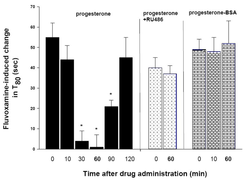
Time course for local application of progesterone alone to inhibit the ability of fluvoxamine to slow 5-HT clearance (left panel); in combination with the progesterone receptor antagonist, RU486 (middle panel); or progesterone-BSA conjugate’s inability to alter the effect of fluvoxamine (right panel). RU486 (1.2 nmoles) was given 15 min before the application of P (0.4 nmoles). The ability of fluvoxamine to increase the T80 value was examined at time 0 (before application of P, P+RU486 or P-BSA) and after their application. Bars and brackets represent the increase in the T80 value, in seconds, after fluvoxamine ± SEM (n=6-8). The effect of fluvoxamine was statistically different at different times post-progesterone administration, as shown by one way ANOVA (F(5,45)=14.180, p<0.001). *p<0.002, Student-Newman-Keuls post hoc analysis comparing the fluvoxamine-induced increase in the T80 value after progesterone at each time point with the value before progesterone administration. RU486 prevented the inhibitory effect of progesterone and P-BSA did not mimic the inhibitory effect of progesterone.
DISCUSSION
These results demonstrate that behaviorally, in OVX rats treated with either EB or P or their combination, fluvoxamine has a markedly reduced ability to induce an antidepressant-like effect in the FST, consistent with these hormones blocking the ability of fluvoxamine to slow 5-HT clearance after similar treatment. Local application of E2 decreases SERT function as the clearance of exogenously applied 5-HT is slower and also inhibits fluvoxamine’s effect on 5-HT clearance, at both early and later time points. Studies with an ER antagonist indicate that this effect of E2 on 5-HT clearance is distinct from its ability to block the inhibitory effect of fluvoxamine on 5-HT clearance. Indeed, studies with ER subtype-selective agonists show that these two effects of E2 are mediated by different ER subtypes. ERβ and GPR30 may play a role in the effects of E2 on basal clearance whereas its blockade of fluvoxamine’s effect on 5-HT clearance is mediated through activation of ERα. Progesterone has a different effect on SERT function as its local application had no effect on 5-HT clearance but it does block the ability of fluvoxamine to slow 5-HT clearance, only at the later time point, and this effect is prevented by blockade of the progesterone receptor.
Differences have been seen between males and females in the FST. Females, at all stages of the estrous cycle, are reported to be less immobile than males in the FST (36), or to be less immobile only during proestrus (37). AD-like effects of estradiol in the FST, similar to those produced by SSRIs, were demonstrated in rats and in mice (11,12). By contrast, the AD-like effect of progesterone was similar to that of selective noradrenergic reuptake inhibitors (i.e. decreased immobility and increased climbing behavior) (13). In the present study, neither estradiol nor progesterone alone or combined, administered once to OVX rats, had an AD-like effect in the FST (Figure 2). It is likely that this difference with the findings cited above could be due to variability in the treatment paradigms used, such as the time elapsed between treatment and the swim test and the duration of hormonal treatment.
Ovarian hormones can act either by a genomic mechanism, using classical nuclear transcription factor receptors requiring at least 30-60 minutes to be manifest and is associated with changes in protein synthesis (38), or by a non-genomic, less well-characterized, rapid (seconds to minutes) membrane receptor signaling mechanism (39-42). Steroid- bovine serum albumin (BSA) conjugates are useful tools to study the membrane-mediated action of steroids since they are biologically active and do not penetrate readily the plasma membrane of the cell (43,44). We used receptor agonists and antagonists, hormone-BSA conjugates and the time course of hormone responses (earlier vs later) to try to elucidate those hormone receptors responsible for the effects observed.
In addition to interfering with the effect of fluvoxamine, estrogen given either systematically (8) or locally had an inhibitory effect of its own on 5-HT clearance (Figures 3 and 4). Although fluvoxamine no longer slowed 5-HT clearance in OVX rats administered estradiol systematically or locally, the inhibitory effect of estradiol alone on 5-HT clearance may have produced a “ceiling effect” so as to compromise any further inhibitory effect of fluvoxamine. However, some of the data presented in Figure 5 argue against this possibility. Most importantly, at 10 min the receptor antagonist, ICI 182,780, restored the inhibitory effect of fluvoxamine even though basal clearance still remained inhibited. This makes it unlikely that the ability of E2 to inhibit the effects of fluvoxamine was due to a “ceiling effect” and indicates that the inhibitory effect of E2 alone on 5-HT clearance is not due precisely to the same mechanism by which it inhibits the effect of fluvoxamine. This was further confirmed by the use of ER subtype-specific agonists (see below). From the time course of E2’s effects, together with those of the E2-BSA conjugate reported previously (8), as well as the effects of ICI 182,780, it may be inferred that E2 has (1) rapid, presumably non-genomic effects, only one of which is mediated by a receptor antagonized by ICI 182,780; and (2) possibly later genomic effects, both of which can be blocked by ICI 182,780. It is not known which ER subtype is responsible for mediating these effects.
E2 has actions at both types of classical nuclear receptors, ERα and ERβ. Some of the regional differences in ERα versus ERβ distribution in brain may confer specific functional effects of E2 acting via these receptor isoforms. ERα is predominant in the ventromedial hypothalamus and is related to E2’s facilitatory actions on female rodent sexual responding; ERβ is predominant in limbic and/or stress-responsive regions of the brain, e.g., hippocampus, bed nucleus of the stria terminalis, and paraventricular nucleus of the hypothalamus (45). Thus, E2 may act to alter affective behaviors of rodents in part through activation of ERβ. Effects of E2 in producing anxiety- and/or depression–like behaviors through activation of the ERβ subtype have been reported using both ERβ knockdown and ERβ activation approaches (12, 46-49). It has also been shown that the AD-like effect of estradiol was mediated by activation of ERβ, rather than ERα (12, 50). Consistent with this, our data indicate that the AD-like effect of E2 (i.e., slower 5-HT clearance) was due to ERβ activation as DPN, its specific agonist, mimicked this effect (figure 6). By contrast, E2’s blockade of fluvoxamine’s ability to slow serotonin clearance was mimicked only by ERα agonists, PPT and THC, and seems to be mediated through this receptor subtype (Figure 7). Although it is established that the AD-like effect of E2 is mediated via ERβ, to our knowledge this is the first report showing that E2’s activation of ERα interferes with the ability of an SSRI to block SERT function.
E2 acts not only at classic nuclear ERs but also at the novel G protein-coupled ER, GPR30. In hypothalamus, GPR30 was detected in the paraventricular nucleus and supraoptic nucleus (51). The expression of GPR30 in rats was observed at both mRNA and protein levels in the hippocampus (52). Recently, GPR30 stimulation was shown to attenuate serotonin receptor signaling in the PVN as demonstrated by reduced oxytocin and ACTH responses, suggesting that GPR30 may play a role in mood disorders (53). The activation of GPR30 by its specific agonist, G1, mimicked the effects of E2 in slowing 5-HT clearance whereas it had no effect on the fluvoxamine-induced blockade of 5-HT clearance (Figures 6 and 7). Thus, the effect of E2 on slowing basal 5-HT clearance involves either ERβ and/or GPR30. With regards to signaling, although the nuclear receptors are known to regulate gene transcription they are also capable of activating second messenger signaling pathways such as mitogen activated protein kinases (MAPK), calcium/calmodulin-dependant protein kinases (CamKII) and cAMP response element-binding proteins (CREB) (54, 55). Future studies will be necessary to clarify which signaling pathway(s) are involved in the observed effects of E2 on SERT function. GPR30 functions as a classic G protein coupled receptor. Studies using breast cancer cells demonstrated E2 signaling through GPR30-mediated activation of MAPK through transactivation of epidermal growth factor (EGF) receptor and stimulation of adenyl cyclase and ERK phosphorylation (56, 57). Whether GPR30 utilizes the same signaling processes in the brain remains to be evaluated.
Progesterone and its receptors play a pivotal role in the regulation of brain activity by its modulation of synthesis and release of neurotransmitters and neuropeptides in response to physiological or pathological stimuli (58, 59). Progesterone altered SERT function in a somewhat different manner than estradiol did. Local application of progesterone mimicked the effect of its systemic administration in that it had no effect on 5-HT clearance (Table 1) whereas it blocked the ability of fluvoxamine to slow 5-HT clearance at later time points (Figure 8). This inhibitory effect of progesterone was blocked by its receptor antagonist, RU486. Further the P-BSA conjugate had no effect on the ability of fluvoxamine to slow clearance (Figure 8). Taken together, these results indicate that the effect of P on fluvoxamine’s ability to slow clearance does not involve membrane PRs but are mediated via activation of intracellular PRs. Progesterone can also influence neuronal activity through its metabolite allopregnanolone, a neurosteroid which acts as an agonist on the GABAA receptor (60-62). However, a direct effect of P on its receptors is suggested by the ability of RU 486 to block the inhibitory effect of P and by the fact that PRs are demonstrated to be expressed in the hippocampus (63-65).
Although acute treatment with estrogen decreased 5-HT clearance in the CA3 region of the hippocampus, it did not alter total (both membrane-bound and intracellular) SERT density as measured by the binding of [3H]cyanoimipramine (8). However, the possibility of changes in membrane distribution of SERT cannot be ruled out. Estrogen-regulated changes in SERT expression have been reported in female macaques and rats brain (66, 67). Beside acting on the transporter, estrogen can act at several levels of the serotonergic system, including effects on its synthesis, receptors and function; this could account for estrogen having beneficial effects on mood that are mediated, at least in part, by interaction with serotonergic systems (68-74).
Estrogen enhancement of plasticity is evidenced by increases in neurogenesis, neural network connectivity and synaptic transmission (75). Hormonal (or estrogen alone) therapy has also been reported to have antidepressant effects in perimenopausal and postmenopausal women (5), although this is not a universal finding (6). Of specific importance for this study is the effect of addition of ovarian hormones, particularly estrogen, on the efficacy of SSRIs in patients. The results of such studies are quite variable. There is certainly no consistent body of data demonstrating that elderly depressed women taking estrogen have a better antidepressant response to SSRIs than those not taking the hormone (76-80). Our experiments do not mimic the clinical situation as we are studying the acute interaction of the ovarian hormones with SSRIs. However, the results obtained in our studies in OVX rats show that, when given acutely, estrogen (or progesterone) inhibited the ability of SSRIs to block the serotonin transporter, which could result in a diminution of SSRI efficacy. This effect of estradiol was mediated through ERα activation. By contrast, estrogen’s blockade of 5-HT clearance, which may result in some beneficial enhancement of serotonin function, appears to be a consequence of ERβ and/or GPR30 stimulation. Thus, understanding the mechanisms behind such effects might reveal a strategy, such as targeting of ERβ or GPR30, to permit beneficial effects of estrogen without its deleterious effect on SSRI-induced efficacy.
Acknowledgments
This research was supported by funds from NARSAD to SB, the Department of Veterans Affairs and the National Institute of Mental Health (MH090386) to AF.
Footnotes
FINANCIAL DISCLOSURES: Dr. Benmansour, Ms. Weaver, Ms. Barton and Mr. Adeniji have no biomedical financial interests or potential conflicts of interest.
Dr. Frazer has been on advisory boards for Cyberonics, Inc., H. Lundbeck A/S and Takeda Pharmaceuticals America, Inc. and he has consulted and/or received research support for preclinical studies from Forest Research Institute, Eli Lilly and Company, Wyeth Pharmaceuticals, and H. Lundbeck A/S. No support for this study was received from any pharmaceutical company.
Publisher's Disclaimer: This is a PDF file of an unedited manuscript that has been accepted for publication. As a service to our customers we are providing this early version of the manuscript. The manuscript will undergo copyediting, typesetting, and review of the resulting proof before it is published in its final citable form. Please note that during the production process errors may be discovered which could affect the content, and all legal disclaimers that apply to the journal pertain.
References
- 1.Hyde JS, Mezulis AH, Abramson LY. The ABCs of depression: integrating affective, biological, and cognitive models to explain the emergence of the gender difference in depression. Psychol Rev. 2008;115:291–313. doi: 10.1037/0033-295X.115.2.291. [DOI] [PubMed] [Google Scholar]
- 2.Pearlstein TB. Hormones and depression: what are the facts about premenstrual syndrome, menopause, and hormone replacement therapy? Am J Obstet Gynecol. 1995;173:646–653. doi: 10.1016/0002-9378(95)90297-x. [DOI] [PubMed] [Google Scholar]
- 3.Amore M, Di Donato P, Berti A, Palareti A, Chirico C, Papalini A, et al. Sexual and psychological symptoms in the climacteric years. Maturitas. 2007;56:303–311. doi: 10.1016/j.maturitas.2006.09.006. [DOI] [PubMed] [Google Scholar]
- 4.Soares CN. Depression during the menopausal transition: window of vulnerability or continuum of risk? Menopause. 2008;15:207–209. doi: 10.1097/gme.0b013e31816381e9. [DOI] [PubMed] [Google Scholar]
- 5.Frey BN, Lord C, Soares CN. Depression during menopausal transition: a review of treatment strategies and pathophysiological correlates. Menopause Int. 2008;14:123–128. doi: 10.1258/mi.2008.008019. [DOI] [PubMed] [Google Scholar]
- 6.Morrison MF, Kallan MJ, Ten Have T, Katz I, Tweedy K, Battistini M. Lack of efficacy of estradiol for depression in postmenopausal women: a randomized, controlled trial. Biol Psychiatry. 2004;55:406–412. doi: 10.1016/j.biopsych.2003.08.011. [DOI] [PubMed] [Google Scholar]
- 7.Butler SG, Meegan MJ. Recent developments in the design of anti-depressive therapies: targeting the serotonin transporter. Curr Med Chem. 2008;15:1737–1761. doi: 10.2174/092986708784872357. [DOI] [PubMed] [Google Scholar]
- 8.Benmansour S, Piotrowski JP, Altamirano AV, Frazer A. Impact of ovarian hormones on the modulation of the serotonin transporter by fluvoxamine. Neuropsychopharmacology. 2009;34:555–564. doi: 10.1038/npp.2008.23. [DOI] [PubMed] [Google Scholar]
- 9.Porsolt RD, Bertin A, Jalfre M. Behavioral despair in mice: a primary screening test for antidepressants. Arch Int Pharmacodyn Ther. 1977;229:327–336. [PubMed] [Google Scholar]
- 10.Cryan JF, Markou A, Lucki I. Assessing antidepressant activity in rodents: recent developments and future needs. Trends Pharmacol Sci. 2002;23:238–245. doi: 10.1016/s0165-6147(02)02017-5. [DOI] [PubMed] [Google Scholar]
- 11.Estrada-Camarena E, Fernandez-Guasti A, Lopez-Rubalcava C. Antidepressant-like effect of different estrogenic compounds in the forced swimming test. Neuropsychopharmacology. 2003;28:830–838. doi: 10.1038/sj.npp.1300097. [DOI] [PubMed] [Google Scholar]
- 12.Rocha BA, Fleischer R, Schaeffer JM, Rohrer SP, Hickey GJ. 17 Beta-estradiol-induced antidepressant-like effect in the forced swim test is absent in estrogen receptor-beta knockout (BERKO) mice. Psychopharmacology (Berl) 2005;179:637–643. doi: 10.1007/s00213-004-2078-1. [DOI] [PubMed] [Google Scholar]
- 13.Molina-Hernandez M, Tellez-Alcantara NP. Antidepressant-like actions of pregnancy, and progesterone in Wistar rats forced to swim. Psychoneuroendocrinology. 2001;26:479–491. doi: 10.1016/s0306-4530(01)00007-5. [DOI] [PubMed] [Google Scholar]
- 14.Kuiper GG, Enmark E, Pelto-Huikko M, Nilsson S, Gustafsson JA. Cloning of a novel receptor expressed in rat prostate and ovary. Proc Natl Acad Sci U S A. 1996;93:5925–5930. doi: 10.1073/pnas.93.12.5925. [DOI] [PMC free article] [PubMed] [Google Scholar]
- 15.Green S, Walter P, Greene G, Krust A, Goffin C, Jensen E, et al. Cloning of the human oestrogen receptor cDNA. J Steroid Biochem. 1986;24:77–83. doi: 10.1016/0022-4731(86)90035-x. [DOI] [PubMed] [Google Scholar]
- 16.Carmeci C, Thompson DA, Ring HZ, Francke U, Weigel RJ. Identification of a gene (GPR30) with homology to the G-protein-coupled receptor superfamily associated with estrogen receptor expression in breast cancer. Genomics. 1997;45:607–617. doi: 10.1006/geno.1997.4972. [DOI] [PubMed] [Google Scholar]
- 17.Prossnitz ER, Arterburn JB, Sklar LA. GPR30: A G protein-coupled receptor for estrogen. Mol Cell Endocrinol. 2007;265-266:138–142. doi: 10.1016/j.mce.2006.12.010. [DOI] [PMC free article] [PubMed] [Google Scholar]
- 18.Funakoshi T, Yanai A, Shinoda K, Kawano MM, Mizukami Y. G protein-coupled receptor 30 is an estrogen receptor in the plasma membrane. Biochem Biophys Res Commun. 2006;346:904–910. doi: 10.1016/j.bbrc.2006.05.191. [DOI] [PubMed] [Google Scholar]
- 19.Thomas P, Pang Y, Filardo EJ, Dong J. Identity of an estrogen membrane receptor coupled to a G protein in human breast cancer cells. Endocrinology. 2005;146:624–632. doi: 10.1210/en.2004-1064. [DOI] [PubMed] [Google Scholar]
- 20.Daws LC, Toney GM, Gerhardt GA, Frazer A. In vivo chronoamperometric measures of extracellular serotonin clearance in rat dorsal hippocampus: contribution of serotonin and norepinephrine transporters. J Pharmacol Exp Ther. 1998;286:967–976. [PubMed] [Google Scholar]
- 21.Malberg JE. Implications of adult hippocampal neurogenesis in antidepressant action. J Psychiatry Neurosci. 2004;29:196–205. [PMC free article] [PubMed] [Google Scholar]
- 22.Spencer JL, Waters EM, Romeo RD, Wood GE, Milner TA, McEwen BS. Uncovering the mechanisms of estrogen effects on hippocampal function. Front Neuroendocrinol. 2008;29:219–237. doi: 10.1016/j.yfrne.2007.08.006. [DOI] [PMC free article] [PubMed] [Google Scholar]
- 23.Page ME, Detke MJ, Dalvi A, Kirby LG, Lucki I. Serotonergic mediation of the effects of fluoxetine, but not desipramine, in the rat forced swimming test. Psychopharmacology (Berl) 1999;147:162–167. doi: 10.1007/s002130051156. [DOI] [PubMed] [Google Scholar]
- 24.Cryan JF, Page ME, Lucki I. Noradrenergic lesions differentially alter the antidepressant-like effects of reboxetine in a modified forced swim test. Eur J Pharmacol. 2002;436:197–205. doi: 10.1016/s0014-2999(01)01628-4. [DOI] [PubMed] [Google Scholar]
- 25.Benmansour S, Cecchi M, Morilak DA, Gerhardt GA, Javors MA, Gould GG, et al. Effects of chronic antidepressant treatments on serotonin transporter function, density, and mRNA level. J Neurosci. 1999;19:10494–10501. doi: 10.1523/JNEUROSCI.19-23-10494.1999. [DOI] [PMC free article] [PubMed] [Google Scholar]
- 26.Benmansour S, Owens WA, Cecchi M, Morilak DA, Frazer A. Serotonin clearance in vivo is altered to a greater extent by antidepressantinduced downregulation of the serotonin transporter than by acute blockade of this transporter. J Neurosci. 2002;22:6766–6772. doi: 10.1523/JNEUROSCI.22-15-06766.2002. [DOI] [PMC free article] [PubMed] [Google Scholar]
- 27.Benmansour S, Deltheil T, Piotrowski J, Nicolas L, Reperant C, Gardier AM, et al. Influence of brain-derived neurotrophic factor (BDNF) on serotonin neurotransmission in the hippocampus of adult rodents. Eur J Pharmacol. 2008;587:90–98. doi: 10.1016/j.ejphar.2008.03.048. [DOI] [PubMed] [Google Scholar]
- 28.Paxinos G, Watson C. The Rat Brain in Stereotaxic Coordinates. Academic; New york, NY: 1986. [DOI] [PubMed] [Google Scholar]
- 29.Lund TD, Hinds LR, Handa RJ. The androgen 5alpha-dihydrotestosterone and its metabolite 5alpha-androstan-3beta, 17beta-diol inhibit the hypothalamo-pituitary-adrenal response to stress by acting through estrogen receptor beta-expressing neurons in the hypothalamus. J Neurosci. 2006;26:1448–1456. doi: 10.1523/JNEUROSCI.3777-05.2006. [DOI] [PMC free article] [PubMed] [Google Scholar]
- 30.Prossnitz ER, Barton M. Signaling, physiological functions and clinical relevance of the G protein-coupled estrogen receptor GPER. Prostaglandins Other Lipid Mediat. 2009;89:89–97. doi: 10.1016/j.prostaglandins.2009.05.001. [DOI] [PMC free article] [PubMed] [Google Scholar]
- 31.Xiao L, Jackson LR, Becker JB. The effect of estradiol in the striatum is blocked by ICI 182,780 but not tamoxifen: pharmacological and behavioral evidence. Neuroendocrinology. 2003;77:239–245. doi: 10.1159/000070279. [DOI] [PubMed] [Google Scholar]
- 32.Stauffer SR, Coletta CJ, Tedesco R, Nishiguchi G, Carlson K, Sun J, et al. Pyrazole ligands: structure-affinity/activity relationships and estrogen receptor-alpha-selective agonists. J Med Chem. 2000;43:4934–4947. doi: 10.1021/jm000170m. [DOI] [PubMed] [Google Scholar]
- 33.Sun J, Meyers MJ, Fink BE, Rajendran R, Katzenellenbogen JA, Katzenellenbogen BS. Novel ligands that function as selective estrogens or antiestrogens for estrogen receptor-alpha or estrogen receptor-beta. Endocrinology. 1999;140:800–804. doi: 10.1210/endo.140.2.6480. [DOI] [PubMed] [Google Scholar]
- 34.Bologa CG, Revankar CM, Young SM, Edwards BS, Arterburn JB, Kiselyov AS, et al. Virtual and biomolecular screening converge on a selective agonist for GPR30. Nat Chem Biol. 2006;2:207–212. doi: 10.1038/nchembio775. [DOI] [PubMed] [Google Scholar]
- 35.Baulieu EE. The steroid hormone antagonist RU486. Mechanism at the cellular level and clinical applications. Endocrinol Metab Clin North Am. 1991;20:873–891. [PubMed] [Google Scholar]
- 36.Barros HM, Ferigolo M. Ethopharmacology of imipramine in the forced-swimming test: gender differences. Neurosci Biobehav Rev. 1998;23:279–286. doi: 10.1016/s0149-7634(98)00029-3. [DOI] [PubMed] [Google Scholar]
- 37.Frye CA, Petralia SM, Rhodes ME. Estrous cycle and sex differences in performance on anxiety tasks coincide with increases in hippocampal progesterone and 3alpha,5alpha-THP. Pharmacol Biochem Behav. 2000;67:587–596. doi: 10.1016/s0091-3057(00)00392-0. [DOI] [PubMed] [Google Scholar]
- 38.Katzenellenbogen BS. Mechanisms of action and cross-talk between estrogen receptor and progesterone receptor pathways. J Soc Gynecol Investig. 2000;7:S33–37. doi: 10.1016/s1071-5576(99)00058-1. [DOI] [PubMed] [Google Scholar]
- 39.Levin ER, Pietras RJ. Estrogen receptors outside the nucleus in breast cancer. Breast Cancer Res Treat. 2008;108:351–361. doi: 10.1007/s10549-007-9618-4. [DOI] [PubMed] [Google Scholar]
- 40.Levin ER. Integration of the extranuclear and nuclear actions of estrogen. Mol Endocrinol. 2005;19:1951–1959. doi: 10.1210/me.2004-0390. [DOI] [PMC free article] [PubMed] [Google Scholar]
- 41.Alyea RA, Laurence SE, Kim SH, Katzenellenbogen BS, Katzenellenbogen JA, Watson CS. The roles of membrane estrogen receptor subtypes in modulating dopamine transporters in PC-12 cells. J Neurochem. 2008;106:1525–1533. doi: 10.1111/j.1471-4159.2008.05491.x. [DOI] [PMC free article] [PubMed] [Google Scholar]
- 42.Muchekehu RW, Harvey BJ. Estradiol rapidly induces the translocation and activation of the intermediate conductance calcium activated potassium channel in human eccrine sweat gland cells. Steroids. 2009;74:212–217. doi: 10.1016/j.steroids.2008.10.013. [DOI] [PubMed] [Google Scholar]
- 43.Blackmore PF, Lattanzio FA. Cell surface localization of a novel non-genomic progesterone receptor on the head of human sperm. Biochem Biophys Res Commun. 1991;181:331–336. doi: 10.1016/s0006-291x(05)81422-6. [DOI] [PubMed] [Google Scholar]
- 44.Ramirez VD, Zheng J. Membrane sex-steroid receptors in the brain. Front Neuroendocrinol. 1996;17:402–439. doi: 10.1006/frne.1996.0011. [DOI] [PubMed] [Google Scholar]
- 45.Shughrue PJ, Lane MV, Merchenthaler I. Comparative distribution of estrogen receptor-alpha and -beta mRNA in the rat central nervous system. J Comp Neurol. 1997;388:507–525. doi: 10.1002/(sici)1096-9861(19971201)388:4<507::aid-cne1>3.0.co;2-6. [DOI] [PubMed] [Google Scholar]
- 46.Walf AA, Rhodes ME, Frye CA. Antidepressant effects of ERbeta-selective estrogen receptor modulators in the forced swim test. Pharmacol Biochem Behav. 2004;78:523–529. doi: 10.1016/j.pbb.2004.03.023. [DOI] [PubMed] [Google Scholar]
- 47.Walf AA, Ciriza I, Garcia-Segura LM, Frye CA. Antisense oligodeoxynucleotides for estrogen receptor-beta and alpha attenuate estradiol’s modulation of affective and sexual behavior, respectively. Neuropsychopharmacology. 2008;33:431–440. doi: 10.1038/sj.npp.1301416. [DOI] [PubMed] [Google Scholar]
- 48.Walf AA, Koonce CJ, Frye CA. Estradiol or diarylpropionitrile decrease anxiety-like behavior of wildtype, but not estrogen receptor beta knockout, mice. Behav Neurosci. 2008;122:974–981. doi: 10.1037/a0012749. [DOI] [PMC free article] [PubMed] [Google Scholar]
- 49.Walf AA, Frye CA. ERbeta-selective estrogen receptor modulators produce antianxiety behavior when administered systemically to ovariectomized rats. Neuropsychopharmacology. 2005;30:1598–1609. doi: 10.1038/sj.npp.1300713. [DOI] [PubMed] [Google Scholar]
- 50.Estrada-Camarena E, Fernandez-Guasti A, Lopez-Rubalcava C. Interaction between estrogens and antidepressants in the forced swimming test in rats. Psychopharmacology (Berl) 2004;173:139–145. doi: 10.1007/s00213-003-1707-4. [DOI] [PubMed] [Google Scholar]
- 51.Brailoiu E, Dun SL, Brailoiu GC, Mizuo K, Sklar LA, Oprea TI, et al. Distribution and characterization of estrogen receptor G protein-coupled receptor 30 in the rat central nervous system. J Endocrinol. 2007;193:311–321. doi: 10.1677/JOE-07-0017. [DOI] [PubMed] [Google Scholar]
- 52.Matsuda K, Sakamoto H, Mori H, Hosokawa K, Kawamura A, Itose M, et al. Expression and intracellular distribution of the G protein-coupled receptor 30 in rat hippocampal formation. Neurosci Lett. 2008;441:94–99. doi: 10.1016/j.neulet.2008.05.108. [DOI] [PubMed] [Google Scholar]
- 53.Xu H, Qin S, Carrasco GA, Dai Y, Filardo EJ, Prossnitz ER, et al. Extra-nuclear estrogen receptor GPR30 regulates serotonin function in rat hypothalamus. Neuroscience. 2009;158:1599–1607. doi: 10.1016/j.neuroscience.2008.11.028. [DOI] [PMC free article] [PubMed] [Google Scholar]
- 54.McEwen B. Estrogen actions throughout the brain. Recent Prog Horm Res. 2002;57:357–384. doi: 10.1210/rp.57.1.357. [DOI] [PubMed] [Google Scholar]
- 55.Manavathi B, Kumar R. Steering estrogen signals from the plasma membrane to the nucleus: two sides of the coin. J Cell Physiol. 2006;207:594–604. doi: 10.1002/jcp.20551. [DOI] [PubMed] [Google Scholar]
- 56.Filardo EJ, Quinn JA, Bland KI, Frackelton AR., Jr Estrogen-induced activation of Erk-1 and Erk-2 requires the G protein-coupled receptor homolog, GPR30, and occurs via trans-activation of the epidermal growth factor receptor through release of HB-EGF. Mol Endocrinol. 2000;14:1649–1660. doi: 10.1210/mend.14.10.0532. [DOI] [PubMed] [Google Scholar]
- 57.Filardo EJ, Quinn JA, Frackelton AR, Jr, Bland KI. Estrogen action via the G protein-coupled receptor, GPR30: stimulation of adenylyl cyclase and cAMP-mediated attenuation of the epidermal growth factor receptor-to-MAPK signaling axis. Mol Endocrinol. 2002;16:70–84. doi: 10.1210/mend.16.1.0758. [DOI] [PubMed] [Google Scholar]
- 58.Pluchino N, Luisi M, Lenzi E, Centofanti M, Begliuomini S, Freschi L, et al. Progesterone and progestins: effects on brain, allopregnanolone and beta-endorphin. J Steroid Biochem Mol Biol. 2006;102:205–213. doi: 10.1016/j.jsbmb.2006.09.023. [DOI] [PubMed] [Google Scholar]
- 59.Brinton RD, Thompson RF, Foy MR, Baudry M, Wang J, Finch CE, et al. Progesterone receptors: form and function in brain. Front Neuroendocrinol. 2008;29:313–339. doi: 10.1016/j.yfrne.2008.02.001. [DOI] [PMC free article] [PubMed] [Google Scholar]
- 60.Schumacher M, Coirini H, McEwen BS. Regulation of high-affinity GABAA receptors in the dorsal hippocampus by estradiol and progesterone. Brain Res. 1989;487:178–183. doi: 10.1016/0006-8993(89)90955-4. [DOI] [PubMed] [Google Scholar]
- 61.Foy MR, Akopian G, Thompson RF. Progesterone regulation of synaptic transmission and plasticity in rodent hippocampus. Learn Mem. 2008;15:820–822. doi: 10.1101/lm.1124708. [DOI] [PMC free article] [PubMed] [Google Scholar]
- 62.Auger CJ, Forbes-Lorman RM. Progestin receptor-mediated reduction of anxiety-like behavior in male rats. PLoS One. 2008;3:e3606. doi: 10.1371/journal.pone.0003606. [DOI] [PMC free article] [PubMed] [Google Scholar]
- 63.Guerra-Araiza C, Coyoy-Salgado A, Camacho-Arroyo I. Sex differences in the regulation of progesterone receptor isoforms expression in the rat brain. Brain Res Bull. 2002;59:105–109. doi: 10.1016/s0361-9230(02)00845-6. [DOI] [PubMed] [Google Scholar]
- 64.Guerra-Araiza C, Villamar-Cruz O, Gonzalez-Arenas A, Chavira R, Camacho-Arroyo I. Changes in progesterone receptor isoforms content in the rat brain during the oestrous cycle and after oestradiol and progesterone treatments. J Neuroendocrinol. 2003;15:984–990. doi: 10.1046/j.1365-2826.2003.01088.x. [DOI] [PubMed] [Google Scholar]
- 65.Waters EM, Torres-Reveron A, McEwen BS, Milner TA. Ultrastructural localization of extranuclear progestin receptors in the rat hippocampal formation. J Comp Neurol. 2008;511:34–46. doi: 10.1002/cne.21826. [DOI] [PMC free article] [PubMed] [Google Scholar]
- 66.Sumner BE, Grant KE, Rosie R, Hegele-Hartung C, Fritzemeier KH, Fink G. Raloxifene blocks estradiol induction of the serotonin transporter and 5-hydroxytryptamine2A receptor in female rat brain. Neurosci Lett. 2007;417:95–99. doi: 10.1016/j.neulet.2007.02.039. [DOI] [PubMed] [Google Scholar]
- 67.Smith LJ, Henderson JA, Abell CW, Bethea CL. Effects of ovarian steroids and raloxifene on proteins that synthesize, transport, and degrade serotonin in the raphe region of macaques. Neuropsychopharmacology. 2004;29:2035–2045. doi: 10.1038/sj.npp.1300510. [DOI] [PubMed] [Google Scholar]
- 68.Bethea CL, Pecins-Thompson M, Schutzer WE, Gundlah C, Lu ZN. Ovarian steroids and serotonin neural function. Mol Neurobiol. 1998;18:87–123. doi: 10.1007/BF02914268. [DOI] [PubMed] [Google Scholar]
- 69.Bethea CL, Gundlah C, Mirkes SJ. Ovarian steroid action in the serotonin neural system of macaques. Novartis Found Symp. 2000;230:112–130. doi: 10.1002/0470870818.ch9. discussion 130-113. [DOI] [PubMed] [Google Scholar]
- 70.Rubinow DR, Schmidt PJ, Roca CA. Estrogen-serotonin interactions: implications for affective regulation. Biol Psychiatry. 1998;44:839–850. doi: 10.1016/s0006-3223(98)00162-0. [DOI] [PubMed] [Google Scholar]
- 71.McEwen BS, Alves SE. Estrogen actions in the central nervous system. Endocr Rev. 1999;20:279–307. doi: 10.1210/edrv.20.3.0365. [DOI] [PubMed] [Google Scholar]
- 72.Cyr M, Landry M, Di Paolo T. Modulation by estrogen-receptor directed drugs of 5-hydroxytryptamine-2A receptors in rat brain. Neuropsychopharmacology. 2000;23:69–78. doi: 10.1016/S0893-133X(00)00085-3. [DOI] [PubMed] [Google Scholar]
- 73.Amin Z, Canli T, Epperson CN. Effect of estrogen-serotonin interactions on mood and cognition. Behav Cogn Neurosci Rev. 2005;4:43–58. doi: 10.1177/1534582305277152. [DOI] [PubMed] [Google Scholar]
- 74.Lasiuk GC, Hegadoren KM. The effects of estradiol on central serotonergic systems and its relationship to mood in women. Biol Res Nurs. 2007;9:147–160. doi: 10.1177/1099800407305600. [DOI] [PubMed] [Google Scholar]
- 75.Brinton RD. Estrogen-induced plasticity from cells to circuits: predictions for cognitive function. Trends Pharmacol Sci. 2009;30:212–222. doi: 10.1016/j.tips.2008.12.006. [DOI] [PMC free article] [PubMed] [Google Scholar]
- 76.Huttner RP, Shepherd JE. Gonadal steroids, selective serotonin reuptake inhibitors, and mood disorders in women. Med Clin North Am. 2003;87:1065–1076. doi: 10.1016/s0025-7125(03)00061-0. [DOI] [PubMed] [Google Scholar]
- 77.Schneider LS, Small GW, Clary CM. Estrogen replacement therapy and antidepressant response to sertraline in older depressed women. Am J Geriatr Psychiatry. 2001;9:393–399. [PubMed] [Google Scholar]
- 78.Schneider LS, Small GW, Hamilton SH, Bystritsky A, Nemeroff CB, Meyers BS. Estrogen replacement and response to fluoxetine in a multicenter geriatric depression trial. Fluoxetine Collaborative Study Group. Am J Geriatr Psychiatry. 1997;5:97–106. [PubMed] [Google Scholar]
- 79.Amsterdam J, Garcia-Espana F, Fawcett J, Quitkin F, Reimherr F, Rosenbaum J, et al. Fluoxetine efficacy in menopausal women with and without estrogen replacement. J Affect Disord. 1999;55:11–17. doi: 10.1016/s0165-0327(98)00203-1. [DOI] [PubMed] [Google Scholar]
- 80.Zanardi R, Rossini D, Magri L, Malaguti A, Colombo C, Smeraldi E. Response to SSRIs and role of the hormonal therapy in post-menopausal depression. Eur Neuropsychopharmacol. 2007;17:400–405. doi: 10.1016/j.euroneuro.2006.11.001. [DOI] [PubMed] [Google Scholar]
